
I decided I am not going to bury the lead on this one. Brian Thomas of the Institute for Creation Research (ICR) just posted another in a long line of creationist screeds attacking the evidence for evolution from comparative embryology, which as usual claims that the evidence is based on fraud and pins much of the blame for it on 19th century biologist Ernst Haeckel.
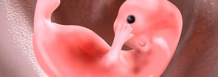
I began writing a rebuttal straight away but then I happened to take a second look at the bright pink image of an embryo atop the article and it brought me to a sudden halt. So, having backed up, let me start again.
Thomas: German zoologist Ernst Haeckel is perhaps most famous for defending evolution with the argument that creatures replay their evolutionary past when developing in the womb. …In his zeal to promote evolution, Haeckel foisted faulty embryo sketches onto his readers, and the zeal of his followers has perpetuated those falsehoods for over a century. (Thomas 2012, emphasis mine)
Yeah, about that…

That’s right, yet another irony meter has been reduced to subatomic particles by a creationist.
It seems that in his zeal to condemn Haeckel and to deny the evidence for evolution from comparative embryology Mr. Thomas has himself foisted a “partially faked” embryo image on his own readers.
As previous ICR authors have done, Mr. Thomas has gone to a stock photography site and purchased an image to illustrate his article, this time one that is ostensibly of a human embryo. However, as soon as I actually looked at the image, I realized it was just, wrong. So I immediately started trying to find where the image had come from, first checking the stock photograph site that I had found the denizens of the ICR using twice before and when that failed to turn up anything, I went, reluctantly, to the Google mine.
Naturally, punching “stock photographs” into Google provided a multitude of links; however, the interweb gods were apparently smiling on me because the very first site I clicked on, iStockphoto, turned up the picture that graces Mr. Thomas’ article. After further investigation I was able to identify the author of the image and was ultimately able to contact him via Google+ (turns out it does have its uses).
Without going into the details, I told him that I needed information about his picture because someone had used it in a scientific context (ha!) and that there were questions about its biological accuracy that I needed to clear up. I asked him about the source of his embryo picture, whether he had created it by manipulating an existing photograph of an embryo or, as I suspected, he had redrawn it from scratch.
The author kindly informed me that he had indeed redrawn the picture based on “several reference images” he had of 7-8 week old embryos. Furthermore, he said that he had not intended to convey biological accuracy with the image and that he had taken “some liberties for artistic and illustration purposes”.
Please note that I did not identify—or link directly to—the author of the image. I did this deliberately, as I do not want to drag him into this more than is necessary. I do not hold him in anyway responsible for ICR/Thomas’ use of the image. He is just someone trying to earn a few extra bucks with his computer art skills. He was very polite and forthcoming with answers to my queries, even offering to send me the original source images if he could find them. I do not intend to impugn his reputation in any way, or negatively affect his business. On the contrary I thank him for his cooperation and hope he does well.
Now that I have the exposition out of the way, without further adieu, here are the two versions of the picture ICR/Mr. Thomas used (the smaller one is from ICR’s main page while the enlarged, cropped, version is from Thomas‘ article page):

Here for comparison is an actual photograph of an 8-week-old (stage 20) human embryo:
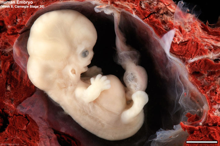
As you can see, it is significantly different from the artistic creation of the iStockphoto author.
Unfortunately, neither Mr. Thomas’ bachelor’s degree in biology nor his master’s in biotechnology seems to have sufficiently prepared him to distinguish between a somewhat fanciful piece of digitally created art and an actual photograph of a biological specimen.
Fear not though, he is absolutely certain that Haeckel’s embryo illustrations alter the true shape of the embryos they depict to such an extent that they create evidence for evolution where none actually exists.
Of course even if Thomas had used the above photograph of an eight-week-old embryo, it would have been fundamentally misleading in this context, because the stages where pharyngeal clefts are evident takes place earlier in development. A more intellectually honest image to have used would be something like this stage 13 human embryo (4-5 weeks old):

In this image not only are the pharyngeal clefts are clearly visible (three left-hand arrows) but the post anal tail is as well (right-hand arrow; something creationist also often deny exists in embryos).
Having dealt with the picture atop the article, I will now return to my rebuttal to the substance of what Mr. Thomas’ wrote, however since I covered the details of pharyngeal clefts pretty thoroughly in “Gill slits” by any other name I will not rehash them here. Instead, I will focus more on Mr. Thomas’ use (abuse) of Haeckel.
Thomas: German zoologist Ernst Haeckel is perhaps most famous for defending evolution with the argument that creatures replay their evolutionary past when developing in the womb. Since Darwin’s time, textbooks have reiterated that early embryos of many vertebrates, including humans, have tiny pouches that reflect an evolutionary fish stage.
Yes, they have but this has nothing to do with Haeckel. It is because of the empirical evidence.
Thomas: More recently, embryologists thoroughly refuted that concept, and others have shown that Haeckel’s drawings were partially faked.
Once again, pharyngeal clefts were first described in amniote embryos by Martin Heinrich Rathke (1793-1860) in 1825 (Rathke 1825a & 1825b). He called them “Schlundspalten” (“throat clefts”) and “Kiemenspalten” (“gill clefts”).
This was nine years before Haeckel was even born!
Creationist scientists—not to be confused with today’s “creation scientists”—regularly called them “gill-slits” and described them as being like the gills of fish during the 34 years intervening their discovery and the publication of Darwin’s On the Origin of Species (1859) which eventually brought evolution into the scientific mainstream (Roget 1834, pp. 631-635) (Carpenter 1839, pp. 320-321) (Agassiz 1849, p. 96).
Most of the illustrations that creationists harp about come from either Haeckel’s 1868Natürliche Schöpfungsgeschichte (The History of Creation) or his 1874Anthropogenie: oder, Entwickelungsgeschichte des Menschen (Anthropogeny: Or, The Evolution of Man).
Did Haeckel develop time travel technology allowing him to send his “partially faked” illustrations back in time so that they could fool all the creationist scientists prior to Darwin publishing? Did he use the same technology to travel into the future to create all of our “faked” embryo photographs as well?
As is typical of creationists when discussing this subject, Mr. Thomas is operating under an implied and unsound syllogism:
I. All evidence for evolution from comparative vertebrate embryology = Haeckel’s drawings & Haeckelian recapitulation.
II. Haeckel’s drawings are, very inaccurate or deliberate frauds and recapitulation has long been discredited.
III. Therefore all evidence from comparative embryology = deliberate fraud and long discredited theory.
Of course given that the premise, in part one, is false, the whole thing falls apart immediately.
Thomas: Embryologist and evolutionist Michael Richardson and colleagues dropped a bomb on Haeckel’s long-held concept known as “embryonic recapitulation.” He compared Haeckel’s old drawings with actual photographs of the same embryos in a 1997 technical paper.1 The comparison showed that Haeckel’s drawings were frauds.
Richardson and his colleagues were using Haeckel as a whipping boy to arguing against the concept of a phylotypic stage in the embryological development of vertebrate embryos. However, when they discovered that creationists were using their work to try to cast doubt on the embryological evidence for evolution—as Mr. Thomas does here—they wrote a letter to the journal Science in response:
A recent study (1) coauthored by several of us and discussed by Elizabeth Pennisi (Research News, 5 Sept. 1997, p. 1435) examined inaccuracies in embryo drawings published last century by Ernst Haeckel. Our work has been used in a nationally televised debate to attack evolutionary theory, and to suggest that evolution cannot explain embryology (2). We strongly disagree with this viewpoint. Data from embryology are fully consistent with Darwinian evolution. Haeckel’s famous drawings are a Creationist cause célèbre (3). Early versions show young embryos looking virtually identical in different vertebrate species. On a fundamental level, Haeckel was correct: All vertebrates develop a similar body plan (consisting of notochord, body segments, pharyngeal pouches, and so forth). This shared developmental program reflects shared evolutionary history. It also fits with overwhelming recent evidence that development in different animals is controlled by common genetic mechanisms (4).
Unfortunately, Haeckel was overzealous. When we compared his drawings with real embryos, we found that he showed many details incorrectly. He did not show significant differences between species, even though his theories allowed for embryonic variation. For example, we found variations in embryonic size, external form, and segment number which he did not show (1). This does not negate Darwinian evolution. On the contrary, the mixture of similarities and differences among vertebrate embryos reflects evolutionary change in developmental mechanisms inherited from a common ancestor (5).
Haeckel’s drawings are used in many modern textbooks, but not always as primary evidence for evolution. In Molecular Biology of the Cell (6), the drawings are used mainly to support hypotheses about the stages of development acted on by natural selection. It is only in this limited context that we have reservations about the implications of the drawings. Thus, certain “phylotypic” embryonic stages, which Haeckel showed as identical, may in fact be significant targets for natural selection.
We are not the first to question the drawings. Haeckel’s past accusers included His (Leipzig University), Rütimeyer (Basel University), and Brass (leader of the Keplerbund group of Protestant scientists). However, these critics did not give persuasive evidence in support of their arguments. We therefore show here a more accurate representation of vertebrate embryos at three arbitrary stages, including the approximate stage (Fig. 1, column three), which Haeckel showed to be identical. We suggest that Haeckel was right to show increasing difference between species as they develop. He was also right to show strong similarities between his earliest embryos of humans and other eutherian mammals (for example, the cat and the bat; Fig. 1, column three). However, he was wrong to imply that there is virtually no evolutionary change in early embryos in the vertebrates (see variations, Fig. 1, column three).
These conclusions are supported in part by comparisons of developmental timing in different vertebrates (7). This work indicates a strong correlation between embryonic developmental sequences in humans and other eutherian mammals, but weak correlation between humans and some “lower” vertebrates. Haeckel‘s inaccuracies damage his credibility, but they do not invalidate the mass of published evidence for Darwinian evolution. Ironically, had Haeckel drawn the embryos accurately, his first two valid points in favor of evolution would have been better demonstrated. (Richardson et al., 1998, emphasis mine)
I have arguments with some of what Richardson et al. say above, but that is for another time. So despite what Mr. Thomas and other creationists might try to imply when they cite Richardson et al. the evidence from comparative embryology is “fully consistent” with evolutionary theory (Haeckel’s problematic illustrations not withstanding).
As for recapitulation, Richardson had this to say about this in a later paper:
Modern views on Haeckel are, typically, ambivalent. His early work is praised, but there is confusion about the Biogenetic Law and recapitulation (the latter being very often confused with embryonic resemblance). Some of this confusion can be blamed on ambiguities and logical flaws in Haeckel’s writing. Several modern studies support the Biogenetic Law in the case of single character transformations.However, there is no evidence from vertebrates that entire stages are recapitulated. Haeckelian and von Baerian models both make the same prediction: that plesiomorphies are transformed into apomorphies during ontogeny. The principle differences between the two models are that Haeckel’s scheme involves heterochrony as one of its mechanisms, and leads to a series of conserved stages. (Richardson & Keuck 2002, p. 522, emphasis mine)
In other words, according to Richardson, there is evidence that some recapitulation does occur, however, it is to a much more limited extent than Haeckel supposedly* advocated. Apparently, something of the structure of recapitulation still stands despite the “bomb” supposedly dropped on it. That is according to Mr. Thomas’ alleged bombardier.
Thomas: Richardson’s report revealed that in order to make animal embryos look more similar at a certain early stage of development, Haeckel had omitted limb buds and heart bulges and resized and selected certain creature’s embryos.2
Some of his criticisms of Haeckel were valid, however others were, to say the least, strained and Richardson has rightfully received some criticisms of his own on this account (Richards 2009).
Please note, though, that Mr. Thomas does not cite Richardson et al.—or anyone else for that matter—as saying that Haeckel added pharyngeal clefts (“gill slits”) that did not exist to the embryos. He talks about limb buds and heart bulges and changes in size of some features, but nothing about “gill slits” which is allegedly the whole point of his article.
Thomas: Since Haeckel had to manipulate data to conform it to his evolutionary notions, then perhaps embryos—including human—do not rehearse their “evolutionary past” after all.
No, Haeckel’s “manipulations” real and imagined are irrelevant. We know that some characteristics of embryological development reflect the phylogeny of the organisms in question and we know this from the collective observations of any number of developmental biologists, none of whom are named Ernst Heinrich Philipp August Haeckel.
For this creationist fantasy to work one must believe the absurdity that no biologist since Haeckel has ever bothered to look at developing embryos and that developmental biology began with Haeckel and ended with him (despite the fact that Haeckel was not himself a developmental biologist).
Thomas: However, textbooks have not yet reflected these findings. For example, the 2007 edition of a popular college biology textbook by Sylvia Mader features a Haeckel-like illustration and an explanation about embryo pouches—sometimes called “gill slits” by evolutionists—and how their presence supposedly supports evolution.
The illustration Thomas is citing (Mader 2007, p. 296) is a bit more than Haeckel-like, in the sense that some of the embryo figures in it look to be redrawn versions of figures that appeared in the 3rd edition of Haeckel’s Anthropogenie (1874).

The figures in green shaded area are clearly redrawn from Haeckel; those in non-shaded portion appear to be based on other sources (see the oldest chick in the Richardson et al. 1998 comparative photo below). The first stage shown for human development in this illustration is technically more accurate than the corresponding stages shown for other animals; however, the second one is less than ideal given that it is of a later stage than that of the corresponding amniote embryos shown.
The primary, legitimate, criticism of these particular figures from Haeckel—and those closely redrawn from them—is that the first stage of embryos shown exaggerates the overall similarity of the embryos it depicts.
Here for comparison is Haeckel’s 1874 first stage drawing of a fish embryo to couple contemporary drawings of fish embryos and a couple photographs:

Haeckel 1874 (plate VI, a generic “fish”), Rathke 1833 (blenny)(reproduced in Richardson & Keuck 2002, p. 510), Deen 1906 (chimaera), Richardson et al. 1998 (dogfish & salmon). Ironically, the pharyngeal clefts of fish embryos (especially more derived fish like Teleosts including the salmon) are often harder to make out than those found in amniote embryos.
And here is Haeckel’s 1874 first stage drawing of a human embryo with similar comparisons:

Haeckel 1874 (plate VII), Haeckel 1910 (plate XII), His 1888 (reproduced on UNSW Embryology website), UNSW Embryology photograph of a stage 12 embryo, 2012 (website).
Richardson et al. (1998) produced the following is a set of comparative photographs of various vertebrate embryos accompanied the letter to Science (reproduced above)
Richardson et al., 1998

I believe that this illustration is problematic as well but it is in the other direction from what is wrong with those by Haeckel. Where Haeckel stands accused, with some justification, of exaggerating the similarities of vertebrate embryos, this set of photographs exaggerates the differences by both the selection of the stages shown and the inclusion of extra embryonic structures (yolk sacs, umbilical stalks etc.). Which, if you do not know what you are looking at can make the embryos seem more fundamentally different than they really are.
But again, that is for another time.
In any case, the figures in Mader (2007), while not photographically accurate do not really create evidence for evolution that is absent in the actual embryos and while updated more technically accurate illustrations would be preferred, illustrations like this one are not the horrible pedagogical malpractice that creationist imply.
This is evident from the fact that creationists rarely address what is specifically wrong with the anatomical details shown in the illustrations that if corrected would contradict evolution. In this article Thomas does mentioned Haeckel’s supposed omission of limb buds & heart bulges, however the omission of these characters do not materially affect their status as illustrations of the evidence for evolution).
Instead, they produce article after article like Mr. Thomas’ which are full of vague accusations about Haeckel having committed fraud in his drawings and implying that all evidence from evolution from developmental biology is suspect with innuendo and guilt by association.
Thomas: Mader wrote:
At some time during development all vertebrates have a postanal tail [spinal cord-like scaffold] and exhibit paired pharyngeal pouches… In humans, the first pair of pouches becomes the tonsils, while the third and fourth pairs become the thymus and parathyroid glands. Why should terrestrial vertebrates develop and then modify such structures like pharyngeal pouches that have lost their original function? The most likely explanation is that fishes are ancestral to other vertebrate groups.3
But how does Mader know that the pouches “lost original function?” She doesn’t—she makes the statement on the basis of evolutionary belief, not on scientific observation. She even lists the pouches’ critical functions for human development. Since the pouches are tissues organized into folds and have known functions, then there is no scientific reason to even suspect that they reflect any evolutionary past.3
Evolution is accepted on the basis of overwhelming evidence, i.e. “scientific observations”, not as a matter of faith, which is what Mr. Thomas is implying. But again, I covered this already in my other post.
The more interesting question here involving lose is how Mr. Thomas lost a significant portion of the paragraph he quotes from Mader. Here is the paragraph as it appears in the original (parts of significance missing in Thomas’ version in boldface):
The homology shared by vertebrates extends to their embryological development (Fig. 17.16). At some time during development, all vertebrates have a postanal tail and exhibit paired pharyngeal pouches. In fishes and amphibian larvae, these pouches develop into functioning gills. In humans, the first pair of pouches becomes the cavity of the middle ear and the auditory tube. The second pair becomes the tonsils, while the third and fourth pairs become the thymus and parathyroid glands. Why should terrestrial vertebrates develop and then modify structures like pharyngeal pouches that have lost their original function? The most likely explanation is that fishes are ancestral to other vertebrate groups. (Mader 2007, p. 296)
I am cynical about the fact that Mr. Thomas replaced the first boldfaced sentence with ellipsis given that it refers to the fact that the pharyngeal apparatus develops into gills, not only in “fish” but in the larvae of amphibians as well but I shall let that pass.
The really striking alteration from the original however are the two boldfaced sentence fragments that follow. This tells the reader that the first set of pharyngeal pouches develops into the middle ear and eustachian tubes in humans and other amniotes (the first pharyngeal cleft develops into the outer ear).
In Mr. Thomas’ version, it incorrectly states that the first pouches develop into the tonsils, skipping the second pair altogether.
I will not speculate on how this omission occurred, by accident or design, it is however a rather significant error. An error, which anyone familiar with the basics of comparative vertebrate embryology, would recognize.
Furthermore, his parenthetical description of the embryonic post anal tail as being a “spinal cord-like scaffold” makes no sense to me.
The spinal cord is the main nerve trunk that extends from the base of the brain, along the vertebral column to the first or second lumbar vertebrae and does not even extend down into the sacral vertebrae—the fused stretch of the spine that joins the pelvic bones—though nerves that branch off it do.
The tail of most tetrapods consists of a number of caudal vertebrae that extend past the sacrum. In humans and other apes, the tail in adults is typically reduced to four caudal vertebrae called the coccyx (though sometimes it is three or five vertebrae).
How Thomas gets to the tail being spinal cord-like—unless he is confusing it with the spine itself—is unclear. It is also unclear how it is supposed to be acting as a scaffold. Though this might be a confused reference to the fact that the coccyx is an attachment point for various muscles, however that is just speculation.
Thomas: A new online video by New Scientist TV, captured by digitally splicing together scans taken during the first trimester of pregnancy, shows the remarkable transformation of these pouches into a human face and head. The pouches show no hint of any loss of function, past or present.
No “hint of any loss” of past function other than the detailed anatomical and genetic similarities between the embryological pharyngeal apparatus of “fish” and amniotes and the fact that the adults of the earliest tetrapods show evidence of having had functioning internal gills.
I am not even sure Mr. Thomas means by “no hint of any loss” of present function. Who is claiming that there is a loss of present function?
Thomas: In fact, the caption accompanying the video states, “A close look at the animation reveals that a face forms from three main features that rotate into place, meeting at the philtrum, the groove above the top lip. The transformation occurs with very precise timing and delays can result in a cleft lip or palate.”4
Very precise timing required an ingenious precision engineer.
Or, it is the result of natural selection. The difference being that natural selection is a well-understood process that has been observed to produce phenotypic changes in populations of living organisms, whereas the existence of Mr. Thomas’ “ingenious precision engineer” is an unobservable theological assertion lacking any scientific support.
Thomas: In his zeal to promote evolution, Haeckel foisted faulty embryo sketches onto his readers, and the zeal of his followers has perpetuated those falsehoods for over a century. Now, not only is his sketch work on embryos “one of the most famous fakes in biology,” but every pouch and fold of the human embryo itself is now known to be fully functional, precisely timed, and intricately arranged.5
The fact that the embryonic pharyngeal apparatus, of humans and other amniotes, has been modified from a structure that develops into fully functional gills to one that develops into other structures that are likewise fully functional is the sort of thing that evolutionary theory predicts.
There is on the other hand no obvious reason that an “ingenious precision engineer” would design such an odd state of affairs.
To expand somewhat on an analogy I used in my previous “gill slit” post, it would be like an engineer starting construction on both a submarine and an automobile by beginning with the same framework. It might technically be possible to do this but it would be far from optimal and make no engineering sense at all.
Evolution on the other hand must work with what it has at hand and in this case it had sarcopterygian fish (submarines) and it modified them into amniotes (automobiles) leaving telltale signs of this history in their embryological development (and in their genes, and in the fossil record).
Thomas: The unaltered data provides accurate information—humans do not rehearse an evolutionary past during embryonic development.
Except when they do.
Thomas: They do rehearse an intricate and expertly designed ballet of precise fit and timing.
Except when they don’t.
Acknowledgements
My thanks to Dr. Andrew “Anj” Petto (University of Wisconsin – Milwaukee), and his contact at McGraw-Hill Higher Education, for getting me a copy of Sylvia Mader’s embryo illustrations (and accompanying text). As well as to my young colleague at Eye on ICR for bringing my attention to Thomas’ article (via a link to my blog, thanks mate). Also my thanks once again to the author of the iStockphoto embryo picture for his help.
Notes
* I say “supposedly” because there is a lot of misinformation and confusion about what Haeckel actually thought. In some cases, this is due to people interpreting what Haeckel wrote through the lens of their own ideas—or through the sparks of an axe they’re grinding—and Haeckel sometimes facilitated these misunderstandings by being vague in his writings. In others because they have simply not bothered to read Haeckel themselves and instead rely what others have said he said.
Recommended reading
“Haeckel’s ABC of evolution and development” (2002) by Michael Richardson & Gerhard Keuck
“Ernst haeckel’s ontogenetic recapitulation: irritation and incentive from 1866 to our time” (2002) by Klaus Sander
“Pictures of Evolution and Charges of Fraud Ernst Haeckel’s Embryological Illustrations” (2006) by Nick Hopwood
“Accuracy in embryo illustrations” (2008) by the National Center for Science Education
“Haeckel’s embryos: fraud not proven” (2009) by Robert J. Richards
References
Agassiz, Louis (1849) Twelve lectures on comparative embryology: delivered before the Lowell institute, in Boston, December and January, 1848-9, Redding & Co.
Carpenter, William B. (1839) Principles of comparative physiology, Intended as an Introduction to the Study of Human Physiology and as a Guide to the Philosophical Pursuit of Natural History, John Churchill, Soho
Dean, Bashford (1906) Chimæroid Fishes And Their Development, Carnegie Institution of Washington, figure 45, plate VII, legend on p. 186
Haeckel, Ernst (1876) The Evolution of Man Vol. I (3rd Edition), H. L. Fowle
Haeckel, Ernst (1910) The Evolution of Man Vol. I (5th Edition), Watts & Co.
Mader, Sylvia S. (2007) Biology (9th ed.), McGraw-Hill
Rathke, Martin H. (1825a) “Kiemen bei Säugetier”, Isis(1825), 747-749;
Rathke, Martin H. (1825b) “Kiemen bei Vögeln”, Isis(1825), 1100-1101.
Richards, Robert J. (2009) “Haeckel’s embryos: fraud not proven“, Biology and Philosophy 24(1):147–154
Richardson et al. (1998) “Haeckel, Embryos and Evolution“, Science 280(5366): 983, 985-6
Richardson, Michael & Keuck, Gerhard (2002) “Haeckel’s ABC of evolution and development“, Biological Reviews, 77: 495-528
Roget, Peter Mark (1834) Animal and Vegetable Physiology Considered with Reference to Natural Theology, Vol. II (Treatise V of The Bridgewater Treatise on the Power Wisdom and Goodness of God as Manifested in the Creation), William Pickering, London
Thomas, Brian (2012) “Do People Have ‘Gill Slits’ in the Womb?“, Institute for Creation Research (website), downloaded on 7-25-2012
UNSW Embryology (2012) University of New South Wales, website, downloaded on 7-25-2012

Okay, this was yet another brilliant and thorough analysis by you, right up there with your gill-slits post a few weeks ago. I’m flabbergasted that, with a mind like yours — with research, analytical, and writing skills like yours, with intellectual integrity like yours, with the self-energizing passion for getting at the specific, accurate, verified truth-whatever-it-may-be that is yours — that you are having, as you announced recently on this blog, “underemployment” issues. I hope very much you can find a situation that gives you the security you clearly richly deserve.
LikeLiked by 1 person
[Blush] Thanks Caroline!
LikeLike
I regret to inform everyone that immediately after reading Caroline52’s comment that Troy’s head exploded Just swelled right up and……eeww. I agree with everything you said, (says proud wife). I would just like to add one thing. I really enjoy his snarky humor.
LikeLike
LikeLike
Very glad to hear he has a proud wife. Also richly deserved. Sorry about the mess, and loss of spousal cranium, hope it was temporary.
LikeLike
So the image was a bit fishy. This is hardly the first time – some time after the fact I found some Czechs arguing on a creationist blog about the picture in this article. The commenters scored the point in the end, and I get the impression that demonstrating such deception is a good first step to showing a creationist that their position has problems.
As for your own article: wow. That is all.
LikeLiked by 1 person
Pingback: What strange beast is this? | Pharyngula
Pingback: What strange beast is this? – Pharyngula
It’s funny how they always attach themselves to the mistakes in evolution theory, like Haeckl’s embryos and the Piltdown man and others without really understanding why or how they are wrong. From this they will judge the whole of evolution to be wrong.
I guess we are guilty of the same thing with them, they constantly make mistakes, yet we never look past these for their corrections and areas of research they got right. Like …
LikeLike
Lovely post. So good to read a longer piece that is as detailed as it is engaging. I’ll be sharing it with my students as a fine example of reasoned critique.
LikeLiked by 1 person
Google now allows you to upload an image to be used as the search item.
It is called Google Images.
LikeLike
If only the anti-evolutionists would read this with an unbiased eye! Thank you for such a wonderful explanation, and one that is easily understood even by those who are not particularly of a scientific bent.
We need more of you!
LikeLiked by 1 person
For future reference, when trying to track down the origins of pictures on the net Tineye.com is your friend.
LikeLike
Hmm, thanks for the tip Sleeper.
LikeLike
Wow, evolution is like magic. It turns gills into ears, tonsils, and glands (and part of the larynx). Calculate the odds…
Granted, it was a bad choice with the stock photo; however, that’s possibly an oversight vs. the intentional use of known fraudulent drawings for decades in school textbooks. Nothing wrong with a little white lie if it wins the argument, right? Perhaps it will, but the argument will still be incorrect.
O’Rahilly and Müller stated that ‘the pharyngeal clefts of vertebrate embryos … are neither gills nor slits’.1 Blechschmidt is more forceful, concluding that ‘the so-called basic law of biogenetics is wrong.” ‘No buts or ifs can mitigate this fact.’ He adds that the gill stage myth is ‘not even a tiny bit correct or correct in a different form…It is totally wrong’. 2 This view is shared by many mainstream embryologists.
in the end, this is probably an argument evolutionists should give up on. But keep right on with it if you insist, it just makes dogmatic evolutionists sound nuts.
1. R. and Müller, F., Human Embryology & Teratology, Wiley- Liss, New York, p. 9, 1992
2.Blechschmidt,E.,TheBeginningsofHumanLife,Springer-Verlag,New York, p. 32, 1977.
LikeLike
As is par for the course, internet creationists spend more time being smug prigs than they do making sure their unwarranted condescension is justified.
“It turns gills into ears…”
The creationist misrepresents evolution AND development in one moronic quip, thus rendering the remaining strident gibberish that much more irrelevant.
As I wrote, as is par for the course.
LikeLike
So I misrepresented something about human development? Are you saying the 1st and 2nd pharyngeal arches don’t contribute to the development of the ear?
http://download.videohelp.com/vitualis/med/Ear_Devt.html
You’ve used the common dogmatic evolutionist tact of complaining that any argument against your belief is “strident gibberish” so that you don’t have to address the actual questions. I assure you my references are from entirely secular scientists in peer reviewed articles.
Please argue from the facts rather than hyperbole.
LikeLike
No, I am saying that ‘turns gills into ears’ is stupid creationist dogma. Pharyngeal arches are not gills, and thus are NOT ‘turned into’ anything.
Please argue from facts and not strident creationist dogmatic gibberish.
LikeLike
derwood, you are correct that Pharyngeal arches are not gills. They never were, never will be, and are not a remnant of a fishy ancestor as the facts I submitted support.
Of course, it would be good if you could present the argument YOU are trying to make and facts supporting that argument, or is your only point of defense to call people names? Please state your case and submit your evidence for whatever point you are trying to make about me or my references.
“…strident creationist dogmatic gibberish” is about the only thing you’ve put forth as evidence so far, which is about as helpful and mature as “I know you are, but what am I”.
LikeLike
How predictable.
What was it Brad? That I added an adjective to the slur that YOU used initially?
You see, Brad, I simply paraphrased the ‘name calling’ that you had used. I guess that was OK when YOU did it, right?
I see that this is perhaps how you’ve decided to try to cover up your obvious naivete re: developmental processes and evolution – hiding behind the standard ‘the mean old Darwinist called me a poopy head’.
But let us look at your ‘facts’:
“They [pharyngeal apparatus] never were, never will be [gills], and are not a remnant of a fishy ancestor as the facts I submitted support.”
And yet you wrote:
“It turns gills into ears, tonsils, and glands (and part of the larynx). ”
And do you REALLY think that there are not hundreds of non-deceptive embryo pictures that could have been used?
Please.
LikeLike
Do you post on JREF at all?
LikeLike
Pingback: Carnival of Evolution 50: The Teaching Edition « Teaching Biology
Pingback: Mendacious creationists won’t fess up | Playing Chess with Pigeons